- July 29, 2013
- Innovation Insights
- Comments : 0
NBIF-backed research provides new insight to cancer using transparent fish
By Shane Fowler, CBC News | link to original article and to watch television story
New Brunswick researchers have, for the first time in the province, transferred genes from one species into those of another, giving scientists a window into how cells develop in more complex creatures.
The researchers at the University of New Brunswick added genes to zebrafish — small fish that reproduce quickly and have transparent skin during their embryonic stage — using a process known as transgenics.
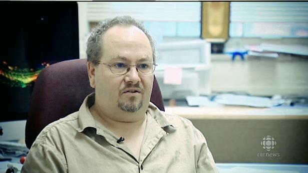
“As far as I know, no one has ever made a transgenic vertebrate animal in New Brunswick,” said Bryan Crawford, the biologist supervising the project.
“Certainly, what we’re doing, nobody has done this before anywhere in the world.”
What Crawford and his team are doing is building a tool set that will allow them to see how cells build, change shape, and migrate through tissues inside a living organism — all without cutting into the creature being studied.
Zebrafish serve as the perfect “lab-rat” for this project because of several traits: one of which is that they’re transparent, which allows for easy viewing of the organisms’ internal development. They also develop rapidly and benefit from having a genome that is relatively easy to manipulate. It also helps that their entire genome has already been sequenced.
If a researcher were to use a lab rat or mouse for this type of project the animal would have to be killed and cut open every time a researcher wanted to check on the cells progression. Not only would that halt cellular development, but also would likely result in the death of the animal.
Emma Chaston-Vickers is the graduate student taking point on the project. She said the extra genes being added to the zebrafish genome allow for the visualization of molecules called Matrix Metalloproteinases, or MMPs.
“I’ve been designing and developing a new technique that will allow us to visualize these molecules in a living embryo,” explained Chaston-Vickers.
“This approach is a little bit different than some past approaches because a lot of the time researchers will isolate these molecules or just study them in cell or tissue cultures, but our approach allows us to look at them in a living embryo.”
MMPs are responsible for, among other things, tearing down the extracellular matrix inside an organism. That matrix is what allows cells to build structures much bigger than themselves. It’s the scaffolding of the cellular world.
The researchers don’t just get to see what’s being built up, but what’s being built wrong — and that’s what is really intriguing, because it allows researchers some insight into how cancer forms.
“When those processes are [not regulated properly] or damaged in some way, you get all kinds of pathologies,” Crawford explains “So like arthritis, tumors, tumour metastasis, cancer, all kinds of things.”
Making the transgenic fish to see what is going on is just the first step.
The next stage of the project will see Crawford and Chaston-Vickers pair with researchers at Dalhousie University to transplant human cancer cells into the transgenic zebrafish.
This will allow them to study the real-time development and growth of cancer, and how the tumor cells break down the extracellular matrix inside a living organism — something that has never been done before.